


Markerless tracking
Tracking the tumor position in X-ray fluoroscopic images
in real time, without metal markers.
In radiotherapy, tumors in the trunk — such as those in the lung and liver — move significantly with breathing. During treatment, the inside of the body is therefore monitored with X-ray fluoroscopic images, and the beam is delivered only when the tumor is at the planned position ("respiratory-gated irradiation").
Conventionally, metal markers are implanted near the tumor to identify its position, but implantation is invasive for the patient. We have developed a technology that tracks the tumor position in X-ray fluoroscopic images in real time without using metal markers. This enables less burdensome treatment for patients and is expected to improve treatment accuracy.
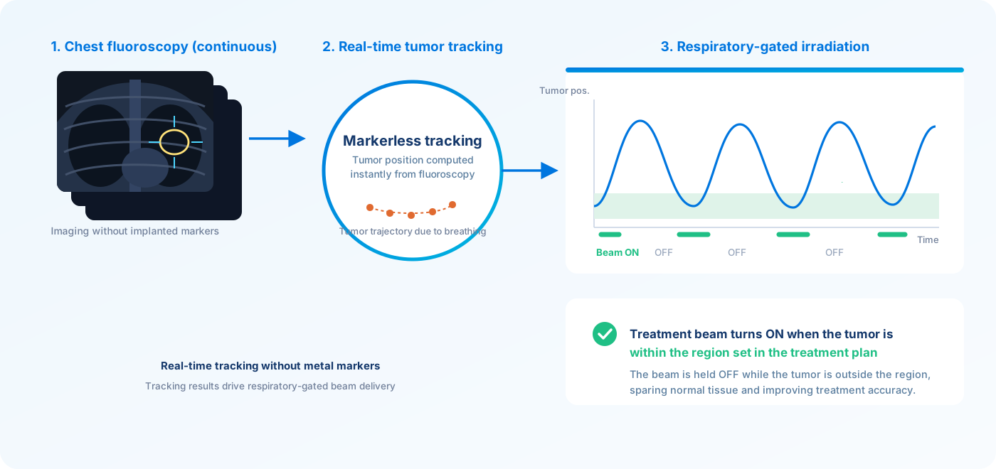
Markerless tracking
Because no metal marker implantation is required, the invasiveness and complication risks associated with implantation are reduced. Hospitalization and preparation for the implantation procedure also become unnecessary, easing the burden on both patients and staff.
Real-time processing
High-speed image processing computes the tumor position instantly from continuously acquired fluoroscopic images. Tracking the tumor without delay as it moves with each breath supports reliable respiratory-gated irradiation.
Improved treatment accuracy
Delivering the beam only while the tumor is within the region set in the treatment plan suppresses unnecessary irradiation of normal tissue. High-precision treatment is achieved even for trunk tumors with large respiratory motion.
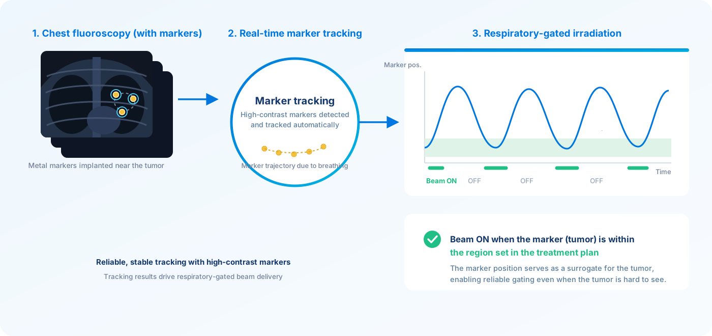
Marker tracking
Tracking metal markers implanted near the tumor
with high accuracy in fluoroscopic images.
Marker tracking is a technique in which metal markers (such as gold markers) implanted near the tumor are automatically detected and tracked in real time in X-ray fluoroscopic images. Because metal markers appear with high contrast in fluoroscopic images, reliable and stable tracking is possible even in regions where anatomical structures overlap in complex ways, or in cases where the tumor itself is difficult to visualize.
The tracked marker position serves as a surrogate for the tumor position and, just as with markerless tracking, enables respiratory-gated irradiation in which the treatment beam is turned ON only while the tumor is within the region set in the treatment plan. We have developed software that supports both markerless tracking and marker tracking, so the optimal method can be chosen according to the clinical case and the facility's workflow.