


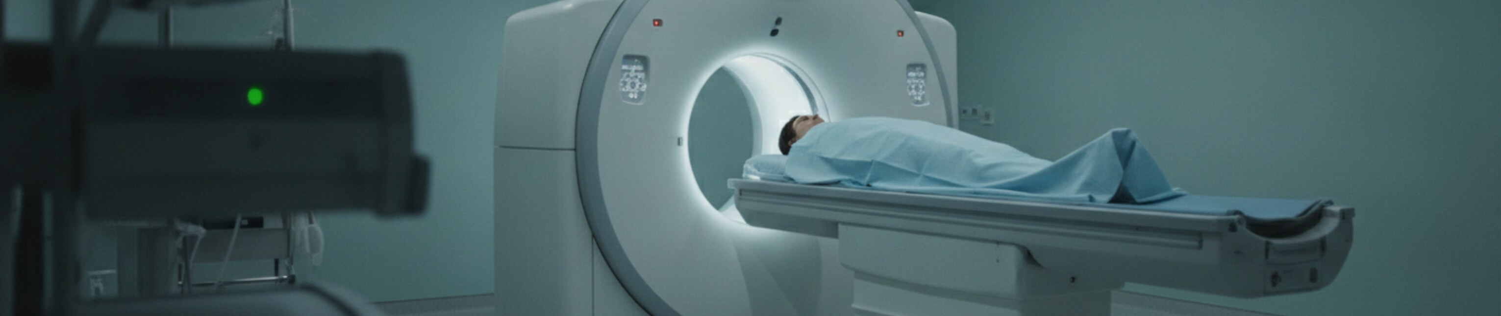
In order to provide cone-beam CT image reconstruction and image processing technology for radiation therapy equipment and particle beam therapy equipment, this product fully utilizes our experience in joint development with diagnostic equipment manufacturers and particle beam therapy equipment manufacturers.
Fast CT image reconstruction
The problem with CT image reconstruction is that it takes a long time to calculate because the calculation cost is high. In the past, multiple FPGAs were used to achieve ultra-high speed, but due to the high development costs, recently it is common to use GPUs for acceleration.

Image processing
Image processing is required for CT images, and both pre-reconstruction data processing and image reconstruction are required to obtain high-quality images. We specialize in data processing before reconstruction, and have implemented various image quality improvement technologies and artifact processing.

Flexible customization
We flexibly respond to joint development with therapeutic device manufacturers and customization according to customer requests. In addition, the introduction of IGRT equipment will enable high-precision treatment even with treatment equipment that does not have CBCT. Please feel free to contact us.
Reconstruction algorithm development
We develop state-of-the-art CT reconstruction algorithms, including iterative reconstruction (IR). Efficient algorithm design and GPU-optimized implementation enable CT image reconstruction in a short time.
Improved spatial resolution
Recently, an X-ray device has been installed in the rotating gantry of a radiotherapy device to obtain CT images (cone beam CT: CBCT). Although the image quality is inferior to that of diagnostic CT images, it has the great advantage of being able to acquire 3D images of the treatment position/condition. However, since linacs and particle beam rotating gantry systems are larger than diagnostic CT systems, the position of the X-ray tube and detector changes during rotation, and the sensitivity of the detector is low.
Ring artifact correction
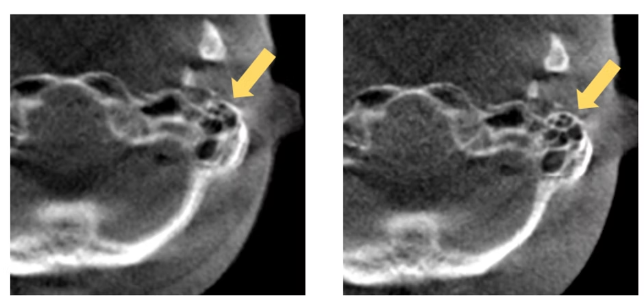
If the sensitivity correction of the detector is not successful, ring artifacts (yellow arrows) will occur in the CT image. In the case of CBCT, the position of the X-ray machine shifts with each gantry angle, so the ring artifacts are not perfectly concentric and cannot be corrected easily. By processing the image before reconstruction, it is possible to correct artifacts with higher accuracy.
CBCT artifact
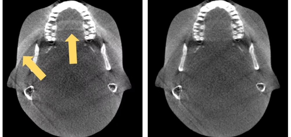
CBCT has the problem that correct image reconstruction is more difficult as it approaches the end in the body axis direction. This is because the X-rays are emitted in a cone shape, so the front area on the detector side is irradiated with X-rays, but the irradiation area becomes narrower as it approaches the X-ray tube side (missing area). On the other hand, several literatures have proposed methods to correct the missing regions. This product performs artifact correction by effectively using the data remaining in the missing area.
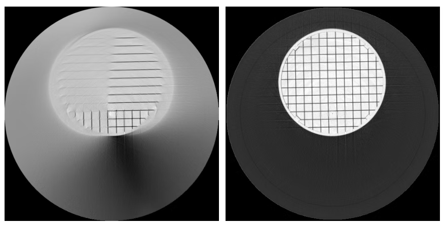
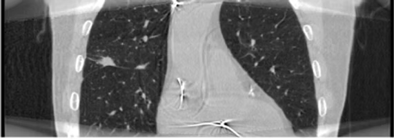
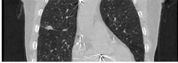
(Left) Before image processing. There are missing areas at the top and bottom edges of the image, and the image is not reconstructed correctly. (Right) After image processing. By effectively using the data of the missing region, CT image reconstruction with reduced CBCT artifacts has become possible.
Phys. Med. Biol. 51 (2006) 3953–39654D-CBCT image reconstruction
Four-dimensional CT (4DCT) captures respiratory motion as a series of CT images along the time axis. The basic concept is to reconstruct each image using only the projection data belonging to the same respiratory phase. However, CBCT acquired on a rotating-gantry treatment machine rotates slowly, making it difficult to capture sufficient respiratory information, and the number of projections is limited. Selecting only the projections of the same respiratory phase reduces the usable data even further, leading to degraded image quality. In other words, patients receive the radiation dose of the CBCT scan, yet not all of it contributes to image quality.
We reconstruct 4D-CBCT images for each respiratory phase using all of the acquired projection data, and combine this with our proprietary image processing to achieve excellent image quality.